Tuesday 16 April 2013

Introduction

Listeria is an eubacteria that is present is 8 forms. As humans, we are most familiar with the form of listeria called Listeria monocytogenes which is the cause of the food borne illness we know as listeriosis. It is the form of listeria that is addressed the most in this blog.

We hear stories about listeria outbreaks in our news sometimes, the most recent one being Manda Packing Company, where many types of meats were recalled due to suspicion of listeria. Many people are not aware of the dangers of listeria, so with careless preparation of food there are chances of contracting listeriosis.

Taxonomy

Below is the taxonomy of listeria as well as the reasons it is categorized in each level of classification. In the brackets are characteristics of listeria that match the characteristics of each level of classification.

Domain: Bacteria (Prokaryotic and no nuclear membrane, unlike eukarya. Has peptidoglycans in cell wall, unlike archea.)

Kingdom: Eubacteria (Has cell wall but no chloroplast, mitochondria, or other organelles.)

Phylum: Firmicutes (It's cell wall is gram positive and it is able to survive harsh conditions.)

Class: Bacilli (Listeria is rod shaped and is able to release endospores)

Order: Bacillales 

Family: Listeriaceae

Genus: Listeria

Species: (depends on type of listeria)
L. monocytogenes
L. grayi
L. innocua
L. seeligeri
L. murrayi
L. ivanovii
L. welshimeri

Life Cycle

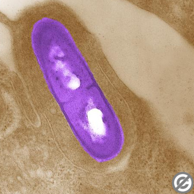
1. Listeria is found in contaminated food.

2. The listeria bacteria is ingested.

3. The bacteria leaves the digestive system and infects the cells in the epithelial lining.

4. The macrophages (cells that aid in removing dead cells and bacteria) in the immune system try to attack and destroy the bacteria through phagocytosis (when the cell engulfs bacteria and digest the bacteria in order to get rid of it).

5. The listeria bacteria is able to get into the cytoplasm and dodge the macrophages because of the enzymes it creates. This enzyme/protien is called listeriolysin O, it is secreted into the macrophages vacuole which damages the macrophages. 

6. Through binary fission, the bacteria is able to reproduce asexually and create identical listeria bacteria in the host cell's cytoplasm.

7. The bacteria use the protein of the host cell to create an actin (a protein found in epithelial and muscle cells) tail in order to transport itself throughout the cells cytoplasm. It uses the tail to propel itself for travel.

8. It creates a filopod (projections of cytoplasm ) by pushing against the cell's outer membrane.

9. It leaves the host cell when the cell comes in contact with a neighbouring cell. It moves into the  neighbouring cell where it repeats the process.

Illistration showing the process of the listeria life cycle.
Shows entering the cell's cytoplasm, binary fission, and leaving the cell's cytoplasm to a neighboring cell
and restarting the process.
From http://www.jhu.edu/cmml/emph_ListSteps.html

Habitat

The listeria bacteria’s natural habitat is in sewage, soil, water, vegetation, food processing plants, and human and animal feces. Listeria is found all over the world because all these habitats are present throughout each continent.  It is also unique because it is able to survive harsh conditions such as heat, high pH levels, and salt levels, which many bacteria are not able to do. It has the capability to survive freezing and refrigeration which is why it is dangerous to humans.

Adaptations

The bacteria is able to adapt and hide inside the host cells cytoplasm and this causes the host’s immune system to be unaware of the bacteria. It is also able to adapt to extremely cold conditions and is able to survive and multiply in those situations. Many bacteria are not able to adapt to cold conditions are do not survive. It also has made an adaption for escaping the immune system’s macrophages. Where is secretes an enzyme that allows it to surpass the macrophages and go into the cell’s cytoplasm.

Another important adaption is the ability to create endospores. Endospores are not like regular reproductive spores, instead they are dormant reproductive cells. They are created when the listeria isn't able to obtain enough nutrients or is in unfavourable conditions. Inside the endospore is a core of DNA and it is able to survive long amounts of time without nutrients. Eventually, when the conditions are favourable, the spore is able to germinate and go back to being a functioning bacteria.

An example of an endospore. (Note that this is an endospore of  Carboxydothermus hydrogenoformans)
From http://www.plosgenetics.org/article/info:doi/10.1371/journal.pgen.0010065

Nutrition

When found in its natural habitat/in nature listeria feeds of dead plants in the sewage or soil that it is living in. This means that listeria is heterotrophic (consumes organisms and organic matter from its surroundings and environment). Once inside the infected host it uses the host cell to obtain it’s nutrients.

Structure and Function

Listeria is a rod shaped bacteria but it can also be found in the form chains of rod shaped bacteria. They are prokaryotic eubacteria (single celled) and contain a plasma membrane, cell wall, ribosomes, and nucleic acid. The bacteria is found to be 0.8-2.6 nanometers in length and 0.6-0.9 nanometers in diameter. They do not reproduce through spores and are non photosynthetic, instead it reproduces through binary fission.
An example of listeria in it's rod shape
From http://www.kimicontrol.com/microorg/Listeria%20monocytogenes.jpg


Listeria is also known to be gram positive, which means that when stained during a gram test the result is a violet or blue – instead of pink or red (gram negative). This shows that the bacteria is likely to not have a thick outer cell wall membrane but has a thick layer of peptidoglycan (a layer around the plasma membrane made of amino acids and sugars) in the cell wall.
Listeria stained during a gram test. Showing violet color therefore gram positive.
From http://coproweb.free.fr/pagbac/introbac/listeria.htm

They move about using flagellum at room temperature (24 degrees) or actin tails provided from host cells at body temperature (37 degrees).